- Issues
- About the Journal
- News
- Cooperation
- Contact Info
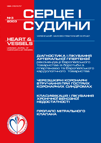
Issue. Articles
№3(3) // 2003

1.
|
Notice: Undefined index: pict in /home/vitapol/heartandvessels.vitapol.com.ua/en/svizhij_nomer.php on line 74
|
|---|
The aim of American National Committee on Prevention, Diagnosis and Treatment of Elevated Blood Pressure (BP) Guidelines and European Society of Cardiology Guidelines for the management of arterial hypertension (AH) is optimization of diagnosis, approaches to the treatment and prevention of AH. According to both Guidelines BP of 140/90 mm Hg and more is elevated. For BP level of 120—139/80—89 mm Hg the new term "prehypertension" was brought into the American Guidelines. Such new risk factors as microalbuminuria and impairment (decreasing) of glomerular filtration were included in the 2003 Guidelines. American Guidelines recommend to start the treatment with the combination on basis of diuretics (thiazides). European Guidelines make physician's choice free, but high efficacy of diuretics and ACE inhibitors' combination, and possibility of diuretics and other antihypertensive drugs' combination are emphasized.
Keywords: arterial hypertension, diagnosis, treatment.
Notice: Undefined variable: lang_long in /home/vitapol/heartandvessels.vitapol.com.ua/en/svizhij_nomer.php on line 143
2.
|
Notice: Undefined index: pict in /home/vitapol/heartandvessels.vitapol.com.ua/en/svizhij_nomer.php on line 74
|
|---|
The most commonly used terms and classifications of chronic diseases of the deep veins of the lower extremities have been analyzed. Their standartization and unified classification is necessary in order to standartize treatment strategies and to evaluate their efficacy and patients' working capacity, as well as the prognosis of the disease. The following terms have been suggested:
- varicous disease of the lower extremities — the disease that primarily affects superficial veins;
- thrombophlebitis — the disease that originates from the inflammation and thrombus formation in superficial veins;
- post-thrombothic disease of the veins of the lower extremities — the disease that originates from deep vein thrombosis.
In order to clarify and standartize the definition of pathological process we suggest that international classification CEAP
should be used in clinical practice. In everyday practice simplified classifications approved in Russia may also be used.
Keywords: classification, chronic venous insufficiency, varicous disease, postGthrombothic disease of the veins, lower extremities.
Notice: Undefined variable: lang_long in /home/vitapol/heartandvessels.vitapol.com.ua/en/svizhij_nomer.php on line 143
3.
|
Notice: Undefined index: pict in /home/vitapol/heartandvessels.vitapol.com.ua/en/svizhij_nomer.php on line 74
|
|---|
Purpose. To improve the medical aid to patients with unstable angina and occlusion of the left main coronary artery.
Materials and methods. 15 patients with unstable angina and occlusion of the left main coronary artery underwent endovascular treatment. Direct stenting, as well as the stenting in combination with predilation was carried out. Intra-aortic ballon counterpulsation was used in case of acute heart failure during or after intervention, as wel as for prevention of heart failure when the bifurcation of the left main coronary artery was affected.
Results. Endovascular intervention was performed to 15 patients suffering from unstable angina with the stenosis of left main coronary artery. 6 patients underwent "pure" angioplasty and 9 (60 %) — coronary stenting that was successful in 100 % of cases. 13 patients underwent emergent percutaneous coronary intervention. PCI was used as a final method of treatment in 12 patients. In 9 cases the median third of left main coronary artery was affected, and 5 of these patients underwent direct stenting. In the other cases the predilation was used. Intra-aortic ballon counterpulsation was used in 5 patients in whom PCI was complicated by acute heart failure, and in order to prevent its development in 2 patients who had the stenosis ofbifurcation of the left main coronary artery. Three patients have died: 2 of them from acute thrombosis of a vessel (only angioplasty was used in these patients), and 1 — from severe myocardial infarction in the pool of the anterior interventricular artery due to incomplete revascularizationthat was performed in order to stabilize patient's status. After PCI 5 patients underwent coronary artery bypass graft surgery — two after "pure" angioplasty, due to restenosis by month 6 and 12, three — due to occlusion of the trunk and other coronal arteries. In other patients episodes of an angina have disappeared, or their frequency and intensity have considerably decreased, which allowed these patients to return to daily work.
Conclusions. Percutaneous coronary angioplasty of the left main coronary artery is an effective method of the treatment. It ensures quick revascularization in patients with unstable angina and may become either final variant of treatment, or a stage of stabilization. During percutaneous coronary interventions in case of stenosis of the left main coronary artery in patients with unstable angina stenting of the affected vessel is essential. When the bifurcation of the left main coronary artery is affected, preventive intra-aortic ballon counterpulsation should be used.
Keywords: percutaneous coronary interventions, stenosis of the left main coronary artery, unstable angina, intra-aortic ballon counterpulsation.
Notice: Undefined variable: lang_long in /home/vitapol/heartandvessels.vitapol.com.ua/en/svizhij_nomer.php on line 143
4.
|
Notice: Undefined index: pict in /home/vitapol/heartandvessels.vitapol.com.ua/en/svizhij_nomer.php on line 74
|
|---|
Purpose. To study the immediate results and follow-up of different methods of recanalisation of the infarct-related coronary artery.
Materials and methods. 135 patients were studied a year after the occurrence of MI. Primary coronary angioplasty was performed in 107 of them (1st group) during the acute phase of MI, primary coronary stenting was performed in 28 (2nd group). Coronary stenting was supplied with intravenous infusion of the glycoprotein IIb/IIIa receptors inhibitor eptifibatid (integrilin) nad with clopidogrel. We studied the clinical course of the disease during the in-hospital stay and in the follow-up period, functional status of the left ventricle and the coronary reserve.
Results. More favourable clinical course of the disease during the in-hospital stay was observed in the coronary stenting group. All the patients of this group remained alive during the following year, left ventricle ejection fraction and threshold capacity were significantly higher than in the coronary angioplasty group.
Conclusions. Percutaneous coronary intervention is an effective method of the treatment of acute MI. Primary coronary stenting supplied with of glycoprotein IIb/IIIa receptor inhibitors may be recommended as the basic strategy in the treatment of acute MI.
Keywords: Primary PTCA, coronary angioplasty, primary stenting, acute myocardial infarction.
Notice: Undefined variable: lang_long in /home/vitapol/heartandvessels.vitapol.com.ua/en/svizhij_nomer.php on line 143
5.
|
Notice: Undefined index: pict in /home/vitapol/heartandvessels.vitapol.com.ua/en/svizhij_nomer.php on line 74
|
|---|
Objective. Thrombolysis remains the standard therapy for acute myocardial infarction (AMI). However, at some institutions primary angioplasty is favored. The purpose of our study was to determine the best reperfusion strategy for patients with AMI.
Materials and methods. Patients with AMI with ST-elevation less than 24 hours duration were assigned to percutaneous coronary interventions (PCI) (n=92) or to thrombolysis (n=75).
Results. The event rates before discharge from the hospital for the PCI group versus thrombolysis group were as follows: mortality: 3,3 % vs 6,6 % (p>0,05), reinfarction: 0 % vs 8 % (p<0,05), recurrent ischemia: 2,2 % vs 34,8 % (p<0,001), Killip class II on the 2—7 day of AMI: 2,2 % vs 28,0 % (p<0,001), left ventricular aneurysm: 17,8 % vs 33,3 % (p<0,05).
Conclusion. Compared with thrombolytic therapy, treatment of patients with primary PCI at hospitals without on-site cardiac surgery is associated with better clinical short-term outcomes.
Keywords: acute myocardial infarction, percutaneous coronary interventions, thrombolysis.
Notice: Undefined variable: lang_long in /home/vitapol/heartandvessels.vitapol.com.ua/en/svizhij_nomer.php on line 143
6.
|
Notice: Undefined index: pict in /home/vitapol/heartandvessels.vitapol.com.ua/en/svizhij_nomer.php on line 74
|
|---|
Despite the progress in surgical treatment of the patients with pulmonary embolism (PE), most of them remain medical patients. In 80 % of the patients the first stage of treatment, including thrombolitic therapy, has been carried out in therapeutic departments.
Purpose. To evaluate the efficacy of thrombolysis in patients with submassive PE and to optimize the indications for this method.
Materials and methods. We studied 84 patients with submassive PE in the age of 18—84 years (mean age 49±6,4). The duration of disease to the moment of receipt in clinic was 3—15 days (mean duration 6±0,8 days). The systolic pressure in pulmonary artery (SPPA) was 66±4,0 mm hg. PE was diagnosed on the basis of clinical signs, ECG, chest X-ray examination, echocardiography. We used the standard criteria: tachycardia, tachypnoe, cyanosis, tendency to hypotension, amplification of 2nd tone on PA; ECG signs of overload of the right side of the heart, intraventricular conduction disturbance; X-ray signs of poor lung pattern, rise of one domes of diaphragm, signs of the lung athelectasis or pleural changes; echocardiographic signs of pulmonary hypertension (PH) and of right vetnricle dilation. In 74 (88,1 %) cases the diagnosis PE was confirmed by pulmonary angiography and in 10 (11,9 %) — by spiral computer tomography. Besides the tool methods listed above, the examination included hematology, biochemical and coagulographic analyses on day 1, 2, 5 and 7. In 56 patients (66,7 %) we revealed deep vien trhombosis of lower extremity as a source of PE. System thrombolysis by streptokinase (1500000 IU) was carried out in 69 patients and with tissue plasminogen activator (100 mg) — in 15 patients not later than on the 10th day from the beginning of disease (mean of 5 ± 1,7 days). All patients were also given enoxaparin sc or heparin IV during 5 days. aPTT was maintained at the level that 1.5—2 times exceeded the initial one, following the prescription of indirect anticoagulants.
Results. Thrombolysis was effective in 67 patients (87,1 %). The functional status of cardiovascular system improved by 1 or more NYHA class at 67 patients (87,1 %), SPPA decreased by the average of 27,3 %, pulmonary resistance decreased by 31,2 %, cardiac index increased by 27,6 % and Ра02 increased by 28,6 % (all p<0,05). In 10 patients (12,9 %) in whom SPPA remained high and PaO2 was low, surgical desobliteration of pulmonary vessels was conducted additionally, in 2 of them artificial circulacion was used. 7 patients (8,3 %) have died. The positive effect of thrombolysis was associated with smaller duration of disease (6±0,8 days vs 18±1,3 days), smaller frequency of post-thromboembolic syndrome (2,9 % vs 66,6 %) and smaller increase of SPPA (51±2 mm Hg vs 66±7 mm Hg), р<0.05.
Conclusions. In patients with submassive PE systemic thrombolysis administered on the 6±0,8 days has good clinical effect in 87,1 % of cases. The predictors of good outcome are the prescription of thrombolytics not later than 10 days and SPPA < 60 mm Hg (their sensitivity is 94.4 % and specificity 74.5 %). In case of long duration of the disease and of high PH surgical treatment should be preferred.
Keywords: pulmonary embolism, postthromboembolic pulmonary hypertension, thrombolysis, venous thrombosis
Notice: Undefined variable: lang_long in /home/vitapol/heartandvessels.vitapol.com.ua/en/svizhij_nomer.php on line 143
7.
|
Notice: Undefined index: pict in /home/vitapol/heartandvessels.vitapol.com.ua/en/svizhij_nomer.php on line 74
|
|---|
Objective. To evaluate xenobiotics — components of cigarettes' smoke — in the organism by investigating the chemical structure of the most widespread sorts of cigarettes in Ukraine.
Materials and methods. Chemical (elements') compound of four cigarettes sorts ("Magna classic" by JT International Ukraine, "Chesterfield lights", "Bond lights" and "Marlboro lights" by Philip Morris Ukraine) was investigated by method of X-ray-fluorescent spectrometry.
Results. According to the analysis of chemical compound of classic cigarettes' smoke, there were detected calcium (45 %), potassium (42,5 %), sulfur (9,5 %) and chlorine (1,6 %). Light cigarettes' smoke containes less amount (for 35—40 %). The whole part of heavy metals' group is 1,1 % of classic cigarettes and 1,7 % of light ones. Concentration of iron is the highest (57,6 %), the second place belongs to manganese (14,7 %) and strontium (15 %); zinc (7,3 %), copper (3 %), chromium (1,2 %) and lead (0,5 %) follow them.
Conclusion. Chemical elements' concentration in cigarettes of different sorts is approximately the same. In case of 20 classic cigarettes per day smoking organism can get 2 kg of different chemical substances for 20 years. The average amount of heavy metals (lead, cadmium, chromium, manganese, iron, copper, strontium, zinc, nickel) may by 23 g for this period.
Keywords: cigarettes, cigarette smoke, concentration of chemical elements, heavy metals.
Notice: Undefined variable: lang_long in /home/vitapol/heartandvessels.vitapol.com.ua/en/svizhij_nomer.php on line 143
8.
|
Notice: Undefined index: pict in /home/vitapol/heartandvessels.vitapol.com.ua/en/svizhij_nomer.php on line 74
|
|---|
Now pulse pressure is considered the one of the most significant and simple markers of elevated risk of complications in patients with cardiovascular diseases.
Objective. To assess the possibility of use of the pulse pressure as a marker of large arteries stiffening in patients with essential hypertension (EH).
Materials and methods. Pulse pressure (PP) in 364 patients 36—73 y.o. (mean 58,4±3,8 year) with I—II stage and I—III EH degree was determined. According to PP level they were divided into 4 groups (≤45, 46—50, 51—64, ≥65). The aortic stiffness was assessed by aortic pulse wave velocity (PWV) in all patients.
Results. The PWV was increased in PP elevation and in the first group it was 7,5± 0,54, in the 2nd — 9,2± 0,6, in the 3rd — 11,4±0,9, in the 4th — 13,9±1,2 (P1—2,3,4<0,05, P2—3,4<0,05, P3—4<0,05). The strong positive correlation (r=0,91) was found between this indices.
Conclusions. The strong positive correlation exists between the PP and aortic PWV in the patients with EH, which allows to use the level of PP as an informative diagnostic marker of large elastic arteries stiffening in patients with EH.
Keywords: pulse pressure, essential hypertension, arteries, remodeling.
Notice: Undefined variable: lang_long in /home/vitapol/heartandvessels.vitapol.com.ua/en/svizhij_nomer.php on line 143
9.
|
Notice: Undefined index: pict in /home/vitapol/heartandvessels.vitapol.com.ua/en/svizhij_nomer.php on line 74
|
|---|
Purpose. To elaborate indications and contra-indications to surgical revascularization of carotids on the basis of the analysis of cerebral hemodynamics, structural changes of the brain experience of carotid endarteriectomy (CE) in patients with significant neurological deficiency due to ischemic stroke (IS).
Materials and methods. We examined 174 patients with rough neurological defects, 49 (28 %) had stenosis of internal carotid artery (SICA) more than 60 % in the pool of the stroke. 30 patients had SICA 70—99 %, 58 had bilateral occlusion. 54 patients underwent CE. In 34 patients we used the internal shunt for (37±7.0) min.
Results. Cerebral blood volume velocity initially was and increased up to the day 7 and 30 after surgery (cm/sec): ipsylateral middle carotid artery (IMCA) — 61.2±3.3, 91.7±3.1, 92.7±4.6, contralateral ipsylateral middle carotid artery (CMCA) — 84.1±2.8, 4.1±7.3, 90.6±1.8, ipsylateral anterior carotid artery (IACA) 71.3±6.6, 88.4±4.2, 97.0±2.1, contralateral ipsylateral anterior carotid artery (CACA) 94.8±6.1, 84.8±8.1, 80.1±9.2, ipsylateral ocular artery (IOA) 6.2±0.8, 7.2±1.1, 10.4±2.46.
Conclusions. The positive clinical effect was observed in 38,8 % of patients. Severe complication occurred in 2 % of cases. At significant neurological deficiency CE is indicated to all the patients with ipsylateral internal carotid artery (IICA) occlusion more than 50 % with contralateral occlusion of carotid artery, with unilateral SICA stenosis more than 70 %, with bilateral stenosis more than 60 %, SICA stenosis of any degree together with destruction of the plaque, or in case of multilacunar strokes in addition to the basic center in the affected hemisphere of a brain with the degree of ICA stenosis that exceeds 60 %. Contraindications to the surgery: subtotal affection of hemispheres, multilevel affection of intracranial vessels, impossibility of the internal shunt implantation. CE stimulates essential increase of the blood flow in IACA, IMCA, CMCA, CACA, and IOA. This effect increases by 30 day after surgery.
Keywords: significant neurological deficiency, carotid endarterectomy, cerebral blood flow.
Notice: Undefined variable: lang_long in /home/vitapol/heartandvessels.vitapol.com.ua/en/svizhij_nomer.php on line 143
10.
|
Notice: Undefined index: pict in /home/vitapol/heartandvessels.vitapol.com.ua/en/svizhij_nomer.php on line 74
|
|---|
Purpose. To define the qualitative and quantitative characteristics of the Doppler spectrum changes at occlusive-stenotic defeats of the arteries of the lower extremities.
Materials and methods. Colour duplex scanning was performed in 599 patients with occlusive atherosclerosis of abdominal aorta and peripheral arteries. The patients were devided into 5 groups. The 1st group — 170 (30,4 %) patients with occlusion of the main artery and hemodynamically significant stenosis of occlusion of the main collateral branch; the 2nd group — 123 (22,0 %) patients with stenosis of the main artery 50—64 %; the 3rd group — 96 (17,2 %) patients with occlusion of the main artery and collateral blood flow in small branches; the 4th group — 64 (11,4 %) patients with main artery stenosis >65 %; the 5th group — 106 (19,0 %) patients with occlusion of the main artery with blood bypass through main collateral.
Results. Decrease of the artery diameter by 20—25 % and 45—50 % contributes to increase of peak systolic volume (PSV) by (56,7±9,4) % and (97,6±15,4) %, respectively. In the 1st group PSV was (49,2±18,4) % lower than in the control group (p<0,05), and their spectrogram was monophase in the 2nd and the 3rd groups pre-stenothic flow. Showed no significant difference from the control group. In the 4th and 5th groups the changes were seen 10—15 cm proximally towards the place of occlusion. Doppler spectrogram was similar to the collateral blood flow, RI decreased by 0,79±0,11 (p<0,05), PSV increased by (32,9±12,7) % (p<0,05). Characteristics of post-stenotic flow was dependent on the grade of stenosis.
Conclusion. Blood flow pattern in lower extremities' arteries depends on the grade of occlusion, collateral flow and peripheral vascular resistance. Stenosis becomes hemodynamically significant on occlusion of 65 % or more. It's signs are: local acceleration, absence of laminar flow and positive diastolic phase (RI 0,83±0,05).
Keywords: artery, occlusion, stenosis, colour duplex scanning
Notice: Undefined variable: lang_long in /home/vitapol/heartandvessels.vitapol.com.ua/en/svizhij_nomer.php on line 143
11.
|
Notice: Undefined index: pict in /home/vitapol/heartandvessels.vitapol.com.ua/en/svizhij_nomer.php on line 74
|
|---|
The aim of our present study was to elaborate algorithm of detection and examination of brahiocephal arteries stenosis in patients with chronical cerebrovascular insufficiency (CCVI), to determine a degree of a stenosis of these arteries, at which one the main Doppler bloodflow indexes authentically differ from the normal, to learn reserves of a cerebral haemodynamic for patients with (CCVI), different stages of CCVI, to determine the indications to surgical treatment according to the obtained data.
Materials and methods. We have selected 205 patients with CCVI; caused by an atherosclerotic lesion in brahiocephal arteries by the data of ultrasound examination, which one were operated by us later. The mean age was 65,5±12 years and was in limits from 48 till 76 years. The men were 142 (69,3 %), wоmen — 63 (30,7 %). We elaborated new algorithm of detection and examination of brahiocephal arteries stenosis in patients with CCVI.
Results. The main Doppler bloodflow indexes in internal carotid and vertebral arteries authentically changed if stenosis of these ones was 75 % and more. The linear bloodflow speed (LBS) on middle cerebral artery (MCA) authentically decreased in patients with ІІ—ІV stage of CCVI, and the advanced of CCVI the lower LBS meanings was. The cerebrovascular reserve (CVR) was determined by LBS monitoring during 3 minutes finger occlusion of common carotid artery. At І CCVI stage patients had high CVR, at ІІ stage — predominantly mean one (80,4 %), at ІІІ stage in most cases (72,9 %) low, and at IV by stage — prevailed (87,5 %) critical degree. Embologenic plaques we observed at any CCVI stage, plaque did not depend on stage. But in patients with І stage percent of such plaques is detected frequently.
Resume. The algorithm, obtained by us, enables to suspect us the CCVI caused by extracranial artery stenoses in 92,6 % of cases, to determine localisation, a degree of a stenosis of these arteries and morphology of an occlusive atherosclerotic plaque in 98 % of cases and to select patients for an operation. The main Doppler bloodflow indexes in internal carotid and vertebral arteries authentically changed if stenosis of these ones was 75 % and are more, therefore these stenoses must be considered as haemodinamically significant. The assay of a brain CVR during transcranial dopplerography is simple in fulfilment, does not demand additional means and enable to estimate authentically a CVR degree elaborated by us. Degrees based on linear bloodflow speed (LBS) changes on middle cerebral artery (MCA) and clinical data. CVR authentically decreases with increasing of a CCVI degree. The operations must be performed in patients with a 75 % and more (66,8 %) stenosis of extracranial arteries with multiple stenoses of those groundwater arteries (33,2 %) if there is neurologic clinic; with ulceration, thrombosis or calcification of an atherosclerotic plaque (31,2 %) at any stage of CCVI.
Keywords: atherosckerosis, extracranial arteries stenosis, diagnostic, cerebrovascular reserv, indications for surgical treatment.
Notice: Undefined variable: lang_long in /home/vitapol/heartandvessels.vitapol.com.ua/en/svizhij_nomer.php on line 143
12.
|
Notice: Undefined index: pict in /home/vitapol/heartandvessels.vitapol.com.ua/en/svizhij_nomer.php on line 74
|
|---|
Objective. To evaluate condition of vascular endothelium in patients with secondary RS due to systemic diseases of connective tissue (SDCТ), clinical efficacy of nafhtidrophuryl and possibility of it's use for correction of endothelial dysfunction.
Materials and methods. 18 women (21—52 y.o.) with the secondary RS, associated with SDCT were examined. RS duration was 1—8 years. Endothelial function by reactive hyperemia test, changes of blood pressure and pulmonary artery pressure by rheopletysmography and dynamics of RS' clinical features during basic therapy with nafhtidrophuryl (enelbin-retard) in the dose of 300 mg/24h for 4 weeks were assessed.
Results. Endothelial dysfunction was revealed in 16 (88,9 %) patients with secondary RS by reactive hyperemia test. Endothelium-dependant dilation of brachial arteria was decreased by 12,5 % (p<0,05) vs healthy people. Nafhtidrophuryl (300 mg/24h) had positive effect in 83,8 % of patients with RS: 24-hours episodes' reduction (34,1 %, p<0,05), temperature normalization period's reduction (24,1 %, p<0,05). The level of endothelium-dependant vasodilation of brachial artery was increased in nafhtidrophuryl — users vs healthy people (by 11,4 %, p<0,05). The negative impact of the drug on BP and pulmonary artery pressure was absent.
Conclusions. Patients with secondary RS due to SDCT have endothelial vascular dysfunction. The use of nafhtidrophuryl (enelbin-retard) — specific 5-НТ2-receptors' blocker — improves clinical features of RS in 83,3 % of patients.
Keywords: Rainand's syndrome, systemic diseases of connective tissue, endothelial dysfunction, 5-НТ2-receptors' blocker, nafhtidrophuryl.
Notice: Undefined variable: lang_long in /home/vitapol/heartandvessels.vitapol.com.ua/en/svizhij_nomer.php on line 143
13.
|
Notice: Undefined index: pict in /home/vitapol/heartandvessels.vitapol.com.ua/en/svizhij_nomer.php on line 74
|
|---|
Mitral valve prolapse (MVP) is widespread. Frequency of this pathology differs and depends on diagnostic methods and criteria. "Strict" criteria should be used for making the diagnosis, especially in the absence of mixomatous degeneration of cusps. It's very important to use recommendations for secondary prevention of infectious endocarditis. Risk factors of complications should be considered in dynamic observation of children with MVP, first of all, in excercises and sports.
Keywords: mitral valve prolapse, diagnosis, complications, treatment.
Notice: Undefined variable: lang_long in /home/vitapol/heartandvessels.vitapol.com.ua/en/svizhij_nomer.php on line 143
14.
|
Notice: Undefined index: pict in /home/vitapol/heartandvessels.vitapol.com.ua/en/svizhij_nomer.php on line 74
|
|---|
The information about Brugada syndrome in modern literature is not enough. That leads to the delay in the diagnosis and proper treatment of this pathology. Ethiology, clinical and electrocardiographical features of Brugada syndrome are shown. The "golden standard" in diagnosis of this pathology is pharmacological test with blocators of fast cardiomyocites' Na+ channels aymalin, first of all. The only effective method of treatment is implantation of cardiovertor-defibrilator in symptomatic patients.
Keywords: Brugada syndrome, sudden death, syncope, blocators of fast Na+ channels.
Notice: Undefined variable: lang_long in /home/vitapol/heartandvessels.vitapol.com.ua/en/svizhij_nomer.php on line 143
15.
|
Notice: Undefined index: pict in /home/vitapol/heartandvessels.vitapol.com.ua/en/svizhij_nomer.php on line 74
|
|---|
The information about Brugada syndrome in modern literature is not enough. That leads to the delay in the diagnosis and proper treatment of this pathology. Ethiology, clinical and electrocardiographical features of Brugada syndrome are shown. The "golden standard" in diagnosis of this pathology is pharmacological test with blocators of fast cardiomyocites' Na+ channels aymalin, first of all. The only effective method of treatment is implantation of cardiovertor-defibrilator in symptomatic patients.
Keywords: Brugada syndrome, sudden death, syncope, blocators of fast Na+ channels.
Notice: Undefined variable: lang_long in /home/vitapol/heartandvessels.vitapol.com.ua/en/svizhij_nomer.php on line 143
16.
|
Notice: Undefined index: pict in /home/vitapol/heartandvessels.vitapol.com.ua/en/svizhij_nomer.php on line 74
|
|---|
The information about Brugada syndrome in modern literature is not enough. That leads to the delay in the diagnosis and proper treatment of this pathology. Ethiology, clinical and electrocardiographical features of Brugada syndrome are shown. The "golden standard" in diagnosis of this pathology is pharmacological test with blocators of fast cardiomyocites' Na+ channels aymalin, first of all. The only effective method of treatment is implantation of cardiovertor-defibrilator in symptomatic patients.
Keywords: Brugada syndrome, sudden death, syncope, blocators of fast Na+ channels.
Notice: Undefined variable: lang_long in /home/vitapol/heartandvessels.vitapol.com.ua/en/svizhij_nomer.php on line 143
17.
|
Notice: Undefined index: pict in /home/vitapol/heartandvessels.vitapol.com.ua/en/svizhij_nomer.php on line 74
|
|---|
The information about Brugada syndrome in modern literature is not enough. That leads to the delay in the diagnosis and proper treatment of this pathology. Ethiology, clinical and electrocardiographical features of Brugada syndrome are shown. The "golden standard" in diagnosis of this pathology is pharmacological test with blocators of fast cardiomyocites' Na+ channels aymalin, first of all. The only effective method of treatment is implantation of cardiovertor-defibrilator in symptomatic patients.
Keywords: Brugada syndrome, sudden death, syncope, blocators of fast Na+ channels.
Notice: Undefined variable: lang_long in /home/vitapol/heartandvessels.vitapol.com.ua/en/svizhij_nomer.php on line 143
18.
|
Notice: Undefined index: pict in /home/vitapol/heartandvessels.vitapol.com.ua/en/svizhij_nomer.php on line 74
|
|---|
Недостаточная освещенность синдрома Бругада в литературе не позволяет вовремя диагностировать большинство случаев столь опасного заболевания и правильно его лечить. Рассмотрены этиология, клинические и электрокардиографические признаки синдрома Бругада. «Золотым стандартом» его диагностики являются характерные изменения ЭКГ при проведении лекарственного теста с применением блокаторов быстрых Na+-каналов кардиомиоцитов, прежде всего аймалина. Единственным эффективным методом лечения является имплантация кардиовертера-дефибриллятора в симптоматичных случаях.
Keywords: синдром Бругада, внезапная смерть, синкопальные состояния, блокаторы Na+-каналов.
Notice: Undefined variable: lang_long in /home/vitapol/heartandvessels.vitapol.com.ua/en/svizhij_nomer.php on line 143
Current Issue Highlights
№4(60) // 2017

Features of different phenotypes development worsening kidney function in acute decompencated heart failure depending on the changes in neutrophil gelatinase-associated lipocalin and initial kidney function
K. M. Amosova 1, I. I. Gorda 1, A. B. Bezrodnyi 1, G. V. Mostbauer 1, Yu. V. Rudenko 1, A. V. Sablin 2, N. V. Melnychenko 2, Yu. O. Sychenko 1, I. V. Prudkiy 1&a
Log In
Notice: Undefined variable: err in /home/vitapol/heartandvessels.vitapol.com.ua/blocks/news.php on line 50

